Tips and Tricks for Bitewing X-Rays

Dental Radiographs are usually called x-rays. Doctors use radiographs for many reasons: to find dental structures which are not visible with naked eye, malignant or benign masses, loss of bone, and cavities.A radiographic image is formed by a controlled burst of X-ray radiation which penetrates oral structures at different levels, depending on varying anatomical densities, before striking the film or sensor. Teeth appear lighter because less radiation penetrates them to reach the film. Dental caries, infections and other changes in the bone density, and the periodontal ligament, appear darker because X-rays readily penetrate these less dense structures. Dental restorations (fillings, crowns) may appear lighter or darker, depending on the density of the material.The ammount of X-ray radiation received by a dental patient is normally low (around 0.150 mSv for a full mouth series, according to the American Dental Association website), equivalent to a few days’ worth of background environmental radiation exposure, or similar to the dose received during a cross-country airplane flight (concentrated into one short burst aimed at a small area). Incidental exposure is reduced by the use of a lead shield, lead apron, sometimes with a lead thyroid collar. Technician can reduce dose of x-rays by leaving the room, or behind proper shielding material, when the X-ray source is active.
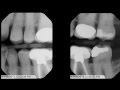
Pacific Dugoni’s radiology department shares tips and tricks for taking bitewing x-rays. www.dental.pacific.edu


No comments: